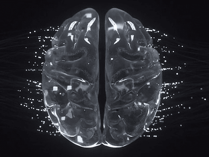
Medical image analysis is the next frontier for deep learning
Medical imaging, including radiography, ultrasound, and MRI, has always required the flexibility of the human eye to detect abnormalities. This is partly because, normally, computers are confused by crowded backgrounds and image quality problems such as specular reflections. However, today, deep learning-based image analysis can automate the search for biological abnormalities in a reliable and repeatable way.
The characteristics of medical images make it incredibly difficult for traditional computer vision algorithms to accurately locate an object or region of interest, particularly for identifying abnormalities in an unstructured scene. It can be time-consuming and difficult, if not impossible, for an automated system to correctly identify the region of interest by ignoring irrelevant features.
Today, deep learning is changing the work of the radiologist, who can now take advantage of computer-aided diagnosis for medical imaging. Whether looking for a specific abnormality, such as a tumor or any deviation from the normal appearance of the body, the Cognex ViDi combines the flexibility of a human specialist’s eye with the speed and robustness of a computer system. In particular, two specialized tools help this process: the ViDi Blue-Locate locates the region of interest, such as a certain organ, even when the background is visually fuzzy or poorly contrasted. The ViDi Red-Analyse tool uses a series of training images to develop a reference model of the normal appearance of that organ, as well as specific types of abnormalities, so that it can mark any shapes that deviate from the normal physiology of the target area as abnormal.
Some interesting examples include the use of deep learning-based tools to locate and identify organs or implants in an x-ray. Cognex ViDi’s Blue-Locate tool can locate a specific organ by learning its distinctive features. To train the Blue-Locate tool, all that is required is to provide images in which the target features are marked. Similarly, deep learning-based defect detection and segmentation tools, such as the ViDi Red-Analyse, can help identify anomalies in the medical image. ViDi Red-Analyse develops reference models of both the normal appearance of an organ and specific abnormalities based on a series of example images. Any abnormalities that deviate from the normal physiology of the affected area are reported to the radiologist, resulting in an automatic assisted diagnosis.


